Meet Todd Clason
Medical researchers’ microscopic imaging needs are highly complex and specialized. Departments at the Larner College of Medicine such as pharmacology and molecular physiology and biophysics rely on research that involves living systems, necessitating tailored and diverse instrumentation.
The Customized Physiology and Imaging Core (CPIC) for the Vermont Center for Cardiovascular and Brain Health (VCCBH) at the college meets the needs of such research, allowing investigators to procure cellular-level images and electrophysiological data that inform research, provide visual evidence, and ideally, confirm findings. The VCCBH also hosts the Molecular Epidemiology and Biostatistics Core, and together, these facilities have supported more than 100 UVM researchers across 90+ projects, leading to 40+ peerreviewed articles since inception in 2020.
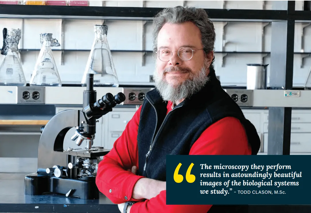
Hired by the anatomy and neurobiology department in 2007 as manager of their imaging facility, Todd Clason, M.Sc., director of the CPIC, has been the keeper of the high-end microscopes and electrophysiology equipment since that time.
Beginning in 2020, the imaging core has been funded under a Centers of Biomedical Research Excellence grant to the VCCBH, co-led by Mary Cushman, M.D., and Mark Nelson, Ph.D. Under the current structure, the Larner community, including graduate students, technicians, and research project leaders, are welcome to use the facility. Clason ensures that potentially intimidating systems in what is known as “Core C”—such as wide-field fluorescence, confocal, spinning disc, and multi-photon microscopes—are accessible to all through consultation and training.
“I train them on what can be imposing pieces of technology and assist researchers to construct a data pipeline that helps them build the data connections they need,” Clason said. “The more people know about the system, the better they’re going to use it.”
Besides providing training and imaging project consultation in the Core, Clason and Doug Taatjes, Ph.D., professor of pathology and laboratory medicine and director of the Microscopy Imaging Center (MIC), teach the Techniques in Microscopy course, which introduces graduate students to all the different kinds of systems that are available in Core C and at the MIC. “Human beings are visual, so if you have data that is visual on your poster or publication, that often engages folks. The microscopy they perform results in astoundingly beautiful images of the biological systems we study,” said Clason.