Medical researchers’ microscopic imaging needs are highly complex and specialized. Departments at the University of Vermont’s Larner College of Medicine such as pharmacology and molecular physiology & biophysics rely on research that involves living systems, necessitating tailored and diverse instrumentation.
The Customized Physiology and Imaging Core (CPIC) for the Vermont Center for Cardiovascular and Brain Health (VCCBH) at the Larner College of Medicine meets the needs of such research, allowing investigators to procure cellular-level images and electrophysiological data that inform research, provide visual evidence, and ideally, confirm findings. The VCCBH also hosts the Molecular Epidemiology and Biostatistics Core, and together, these facilities have supported more than 100 UVM researchers across 90+ projects, leading to 40+ peer-reviewed articles.
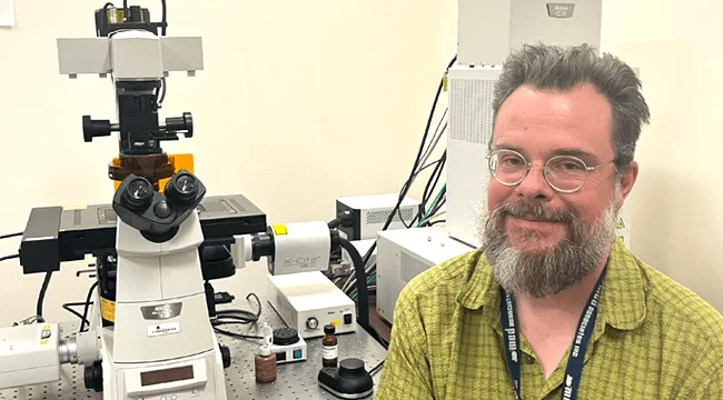
Meet Todd Clason
Hired by the anatomy and neurobiology department in 2007 as manager of their imaging facility, Todd Clason, M.Sc., director of the Customized Physiology and Imaging Core, has been the keeper of the high-end microscopes and electrophysiology equipment since that time.
Clason holds a bachelor of arts in biology and English from Lee Honors College, a master of arts in English language and literature from Western Michigan University, and a master of science in ecology and microscopy from Bowling Green State University.
From 2011 to 2016, Clason was manager of UVM’s imaging facility, renamed the Neuroscience Center of Biomedical Research Excellence under its then director, the late Gary Mawe, Ph.D., Samuel W. Thayer Professor of Neurological Sciences. The facility was fully funded thanks to a COBRE grant awarded to former co-directors and professors emeritus Rodney Parsons, Ph.D., and Cynthia Forehand, Ph.D.
Since 2020, the imaging core has been funded under a COBRE grant to the VCCBH, co-led by Mary Cushman, M.D., and Mark Nelson, Ph.D. Under the current structure, the Larner community, including graduate students, technicians, and research project leaders, are welcome to use the facility. Clason ensures that potentially intimidating systems in what is known as “Core C”—such as wide-field fluorescence, confocal, spinning disc, and multi-photon microscopes—are accessible to all through consultation and training.
“I train them on what can be imposing pieces of technology, and assist researchers to construct a data pipeline that helps them build the data connections they need,” Clason said. “The more people know about the system, the better they’re going to use it.”
Besides providing training and imaging project consultation in the Core, Clason and Doug Taatjes, Ph.D., professor of pathology and laboratory medicine and director of the Microscopy Imaging Center (MIC), teach the Techniques in Microscopy course, which introduces graduate students to all the different kinds of systems that are available in Core C and at the MIC. “Human beings are visual, so if you have data that is visual on your poster or publication, that often engages folks. The microscopy they perform results in astoundingly beautiful images of the biological systems we study,” said Clason.
Life Outside the CPIC
Outside the world of microscopy, Clason and his family enjoy caring for their 14-goat homestead of mostly Nigerian Dwarf goats, and riding around in their vintage 1979 VW bus (when it’s not broken down). “It’s not safe, it’s not fast, but it’s good fun … The bus likes to break down in interesting locations and times,” said Clason. More than two decades ago, they moved from Seattle to Vermont, and eventually to Jericho, where Clason and his wife, Maeve, and their two teenage sons, Phinn and Spencer, reside. In the coming year, Clason’s oldest son plans to study engineering at UVM.
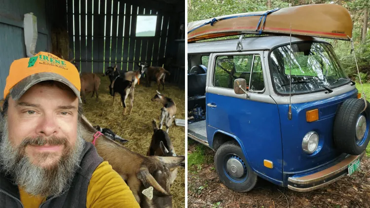
Rural life allows for a garden of currants, blueberries, and fruit trees, not to mention 10 grazing acres for their “spoiled milker” goats with jazzy names like Louis, Lady, Ella, and Duke, who graze outside and sleep in a former horse barn. “We rented goats to see if we could do it. Then we eventually purchased some from New Village Farm in Shelburne, where Spencer went to camp,” said Clason. The family also enjoys a barter economy in their village, where their goat cheese, made mostly by Maeve, is traded with neighbors for eggs.
Imaging Benefits Larner Research
Many past research projects and leaders, particularly those associated with the VCCBH, have been supported by access to the Customized Physiology and Imaging Core, advancing skills in study design, epidemiology, translational research, imaging, experimental instrumentation, and electrophysiology.
Current principal investigators who most frequently use the facility include the Stumpff Lab team under Jason Stumpff, Ph.D., professor of molecular physiology and biophysics who investigate the molecular mechanisms controlling cell division and how they relate to human disease. Research conducted often uses microscopy-based approaches to investigate how chromosomes, which contain the cell’s genetic material, are moved and organized at molecular, cellular, and organismal scales. The images captured in the CPIC help answer such questions as How do kinesin motor proteins contribute to the spatial and temporal control of chromosome movements? and How do defects in these mechanisms contribute to disease? and Can proteins involved in controlling mitotic chromosome movements be exploited as anti-cancer targets?
Similarly, the Nelson Lab team under Mark Nelson, Ph.D., professor and chair of pharmacology, University Distinguished Professor, and co-director of the VCCBH, conducts research on ion channels and calcium signaling in smooth muscle and endothelial cells, including electrical and local calcium signaling in the brain vasculature and urinary bladder. Examples of CPIC-generated imagery for the Nelson Lab include vascular physiology and brain health research related to small vessel disease (SVD)—a major cause of stroke and dementia. Another area of focus that relies on microscopy is bladder function research, related to health issues like overactive bladder and incontinence.
Read more about Dr. Nelson’s Brain Blood Flow Research
More about the CPIC
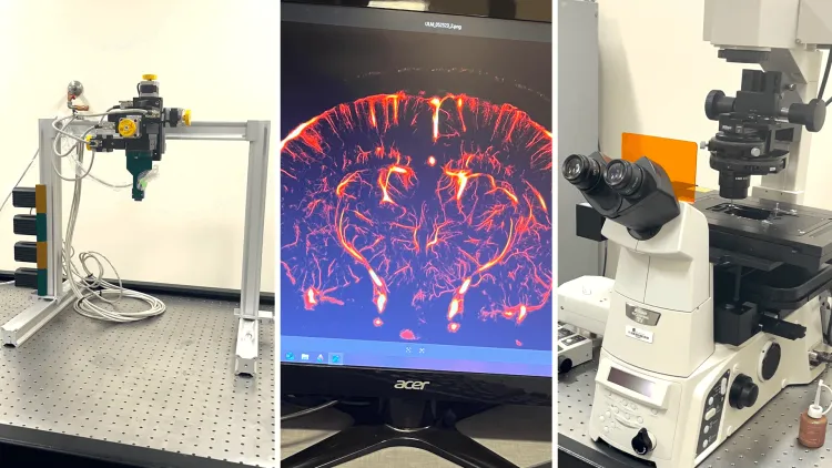
The Customized Physiology and Imaging Core specializes in both pre- and post-experiment assistance, including image analysis, operation of electrophysiology equipment, and the use of 3D printing technologies. Its imaging systems are tailored for studying living systems, such as live animals (in vivo), tissue samples, and cultured cells. The Imaging Core is equipped with a diverse array of instruments, including the following:
- Iconeus One Functional Ultrasound
- Scientifica HyperScope Multiphoton
- Stereo Lithography (SLA) Printer Suite
- Yokogawa CSU-W1 Spinning Disk Confocal Microscope
- Zeiss LSM-7 Multiphoton
- DeltaVision RT Restoration Microscope
- Noran OZ Fast Scanning Confocal Microscopy System
- Nikon C2 Confocal Microscopy System
- Perimed laser Doppler flowmetry system
- Mobile Electrophysiology Instrumentation
Examples of other CPIC services outside of instrumentation training include consultations with Larner pharmacology research assistant professors: electrophysiology with Tom Heppner, Ph.D.; 3D modeling and fabrication with Gerald Herrera, Ph.D.; and image processing and analysis with Grant Hennig, Ph.D.
The facility also supports custom 3D printing for creating specialized live-imaging chambers and experimental devices. Each imaging station can be outfitted with a perfusion system, and electrophysiology tools are available for integrated data collection. CPIC collaborates closely with the Microscopy Imaging Center (MIC), and both facilities use the iLab platform for scheduling access and equipment reservations.
Funding for the support and guidance of investigators from the Customized Physiology and Imaging Core of the Vermont Center for Cardiovascular and Brain Health Funding was provided by P20 GM135007 from the National Institute of General Medical Sciences (NIGMS) of NIH.