With the launch of its new plastinated anatomy lab, UVM joins a select group of New England institutions providing students with access to professionally prepared anatomical specimens.
Developed for UVM’s growing Occupational Therapy (OT) doctoral program, the lab was funded through a federal appropriation secured by former U.S. Senator Patrick Leahy. It gives students the opportunity to study real human anatomy using durable, highly detailed specimens.
“This lab is an amazing resource to understand how the human body functions,” said Victoria Priganc, founding director of Vermont’s first OT program, who led the project.
The specimens are created through plastination, a preservation process that replaces water and fat with polymers, producing odorless, long-lasting models that maintain intricate anatomical detail. Students can examine muscles, nerves, organs, and vessels in three dimensions without needing to learn dissection techniques.
“The plastinated bodies are dissected by professionals, and they’re very intricate, delicate dissections,” Priganc said. “Students can focus on learning anatomy instead of worrying about how to perform a dissection.”
Unlike traditional cadaver labs, plastinated models do not require refrigeration or specialized ventilation and can be used over long periods. For students who do not need surgical training, they offer a practical and accessible alternative.
The collection, valued at approximately $500,000, arrived on campus late last fall. Priganc said it has already changed how students study.
“I often see students back in the lab after class, reviewing their notes and then looking again at the specimens to reinforce what they are learning,” she said.
Occupational therapy students Ella Pratt and Kate Strike say the lab has made a noticeable difference in their understanding.
“You can clearly see structures that are hard to identify in a traditional cadaver lab,” Pratt said. “The models hold their formation.”
Strike, who identifies as a visual learner, said the lab helped her connect lecture material to real anatomy.
“It has helped me understand what is happening in the body, both conceptually and visually,” she said.
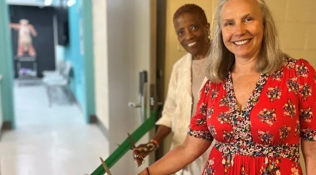
UVM officially unveiled the lab on May 18 during a ribbon-cutting ceremony attended by faculty, students, and supporters.
“It is a pleasure to have everyone here to celebrate this new anatomy lab,” Dean Noma Anderson said. “This marks an important milestone for the college and for UVM.”
Anderson and Priganc opened the space together.
“This is more than a new facility,” Anderson added. “It is a learning environment where future health professionals will build the knowledge, skills, and respect for the human body that will shape their careers.”
She noted that the lab enhances learning across a range of styles and strengthens the connection between classroom instruction and patient care.
“This space supports diverse learners and helps bridge the gap between theory and practice,” Anderson said.
Anderson also recognized the collaborative effort behind the project, including the faculty and staff who contributed to its development, and expressed gratitude for federal and philanthropic support.
“We are deeply grateful to Senator Leahy, as well as the donors and partners who made this possible,” she said.
The plastinated anatomy lab is one of three new learning environments funded through the Leahy appropriation. The others include a pediatric simulation lab and a fully equipped smart apartment. Together, these facilities support UVM’s growing OT program as well as other health sciences students in the College of Nursing and Health Sciences.
“Because of this funding, we now have three advanced teaching labs,” Priganc said. “It has been transformative for our program.”
The students say the investment reflects a strong commitment to their education.
“It shows how much the college values our success,” Strike said.
Pratt agreed. “These models are incredibly valuable,” she said. “They show how committed UVM is to help us succeed.”