
Researchers studying epilepsy at the Larner College of Medicine use a cutting-edge method that received a recent Nobel Prize in Physiology or Medicine. This method, called optogenetics, makes it possible to see how nerve cells shape memories, feelings, and behaviors in the living brain, and to control neural circuits with remarkable precision to manage seizures and cognitive problems.
The 2026 Nobel Prize was awarded to the scientists who developed optogenetics beginning in 2005. Two of the laureates, German biophysicists Peter Hegemann, Ph.D., and Georg Nagel, Ph.D., discovered a light-sensitive protein in a single-celled algae, and a third laureate, Karl Deisseroth, M.D., Ph.D., professor of psychiatry and behavioral sciences at Stanford University, transformed the protein into a light-controlled switch for nerve cells. Since its emergence, optogenetics has transformed how researchers worldwide study brain function and disorders, offering insights into conditions such as epilepsy, depression, and autism, particularly when traditional treatments are ineffective.
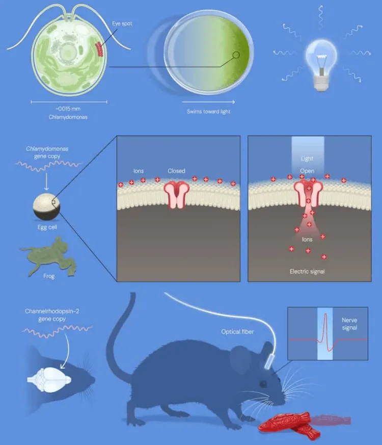
We asked Larner College of Medicine researchers how they use optogenetics technology to advance their own research in Vermont.
Jeremy Barry, Ph.D., Associate Professor of Neurological Sciences
Director of the Epilepsy Cognition and Development Lab at the University of Vermont
Dr. Barry uses optogenetics to study a genetic form of epilepsy caused by changes in a gene called KCNT1. This form of epilepsy causes severe seizures in infants and a frontal lobe epilepsy that often strikes during sleep. Children with these changes often have seizures that don’t respond to existing medications, and many also struggle with learning and memory problems that current treatments barely address. Drawing a line between the neural circuits that both cause seizures and changes in cognition is Dr. Barry’s focus.
“The brain depends on “brake” cells that keep electrical activity in check,” Barry said. “Our work with collaborators at Virginia Polytechnic Institute suggests that the KCNT1 mutation weakens one type of brake cell in the hippocampus, a brain region essential for learning and memory. When those brakes fail, nearby cells fire together in abnormal bursts. These bursts are the small electrical “spikes” seen between seizures, and they can disrupt memory.” This research is funded by the National Institutes of Health.
“Using optogenetics, we made brake cells sensitive to light. Then we drove them with a gentle, steady rhythm, about five pulses per second, a pace the weakened cells can keep up with. The abnormal spikes dropped. When we pushed the cells faster than they could follow, the spikes came back,” he said.
“By pinpointing which cells go wrong, we can help identify treatments aimed at the underlying cause of both seizures and cognitive problems, rather than just the symptoms.” — Jeremy Barry, Ph.D., Associate Professor of Neurological Sciences
“We’re now building a ‘smart’ system that listens to brain activity and delivers a pulse of light within a thousandth of a second of detecting a warning sign. We’ll test whether this can reduce seizures and improve learning and memory over the long term. To do this, we pair optogenetics with ultra-fine recording probes that track activity layer by layer in the brain, and with custom implants we design and 3D-print here at UVM.”
Christa Habela, M.D., associate Professor of neurological sciences
Pediatric Neurologist at Golisano Children’s Hospital at UVM Health
Dr. Habela builds neural circuits from human cells. She starts with stem cells derived from human skin or blood cells, then coaxes them to become specific types of brain and spinal cord cells.
In a study published this year in the journal Cell Reports Methods, Habela and colleagues at Johns Hopkins University built a “brain-to-spinal-cord circuit” on a chip. Human brain cells grow in one tiny chamber and human spinal cord motor cells grow in another. The brain cells send their long connecting fibers through microscopic channels to reach the spinal cord cells, the same way the real pathway that controls movement is wired. This pathway breaks down in diseases like amyotrophic lateral sclerosis (ALS, or Lou Gehrig’s disease), progressive multiple sclerosis, and hereditary spastic paraplegia.
Optogenetics proved that the chip works. The team made brain cells sensitive to light. When they flashed blue light on the brain chamber, the brain cells fired, and a split second later the spinal cord cells responded. That showed the two sides were truly talking to each other. When they blocked the chemical messenger that the cells use to communicate, the spinal response faded.
Habela now uses the same approach to study genetic epilepsies. Because the cells can come from patients themselves, she can grow a child’s own neurons in a dish, use light to probe how their circuits misfire, and test potential treatments on human cells before they ever reach a patient.
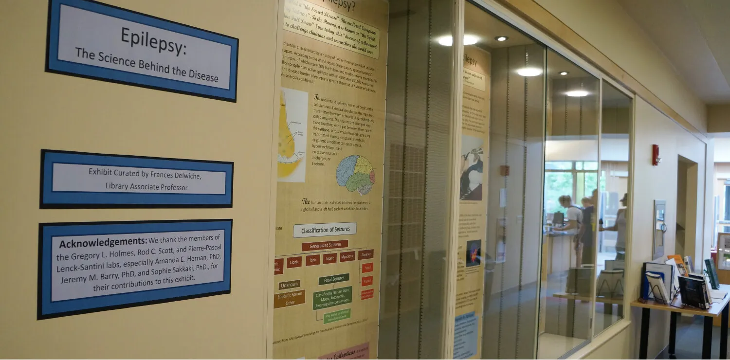
The bigger picture
Barry and Habela are part of a small but growing group at UVM using optogenetics. Molly Stanley, Ph.D., assistant professor of biology and co-director of undergraduate neuroscience in the College of Arts and Sciences, uses optogenetics to directly manipulate and map the neural circuits that govern feeding behaviors and metabolic regulation. By using the fruit fly (Drosophila melanogaster) as a model organism, Dr. Stanley’s lab investigates how specialized taste cells integrate internal energy states to drive dietary choices. By first using the fruit fly whole-brain connectome, pioneered by Davi Bock, Ph.D., associate professor of neurological sciences at the Larner College of Medicine, Stanley’s lab team can make predictions about the function of individual neurons and then use optogenetics to directly activate only those neurons to confirm whether they have the expected outcome on animal behavior.
The primary value of Optogenetics is in discovery, by letting scientists test ideas about what drives a disease and which cells and circuits are malfunctioning. This can points drug developers and gene therapists toward the right targets.
“Optogenetics lets researchers move from asking where a disease lives in the brain to asking which cells cause it, and whether we can fix them.” — Jeremy Barry, Ph.D., Associate Professor of Neurological Sciences
“By building these treatment-testing approaches in established animal models and in cultured patient cells carrying the same gene mutations, we aim to build a world-class program for testing new treatments, from drugs and small molecules to next-generation gene-editing tools.