MRI technology in support of basic science and clinical research
Over 20 UVM research studies currently use the MRI Center for Biomedical Imaging. Besides standard MRI equipment, it includes facilities for delivering experimental stimuli, recording subject responses, acquiring functional images, storing data, and conducting initial image analysis.
MRI Center for Biomedical Imaging Equipment
Overview
The UVM MRI Center for Biomedical Imaging was established and supported in part by funding from the U.S. Department of Energy. Initial funding for the UVM MRI Center was received in 2006. The magnet was installed in April 2007 and operation began in July 2007. The federal grant was renewed in 2008 and again in 2010. In February 2009, the UVM MRI Center was selected by Philips for the first installation in North America of the Achieva 3.0T TX (multitransmit) magnet, one of four such magnets in the world at the time. The MRI Center is owned and operated by the University of Vermont for research purposes and is located in UVM Medical Center's McClure building and is utilized by a broad range of UVM faculty and departments for both basic science and clinical research projects that focus on understanding more about disease and wellness, evaluating new treatments and therapies, and developing new techniques for diagnosis and treatment. More than 20 UVM research studies currently utilize the MRI Center for Biomedical Imaging. In addition to the standard MRI instrumentation, there are additional elements necessary to deliver experimental stimuli, acquire subject responses and functional images, store images, and provide initial image analysis capability. This instrumentation includes both hardware and software.
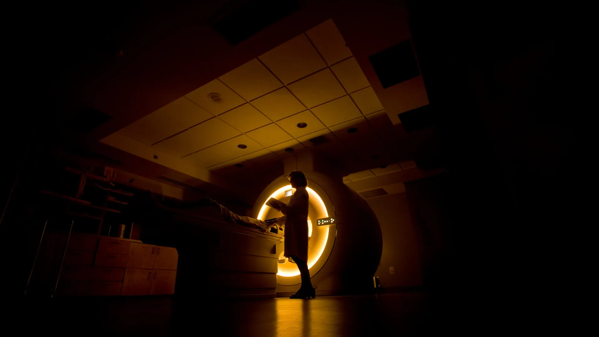
MRI Research Magnet
More than 20 UVM research studies currently utilize the MRI Center for Biomedical Imaging.In addition to the standard MRI instrumentation, there are additional elements necessary to deliver experimental stimuli, acquire subject responses and functional images, store images, and provide initial image analysis capability. This instrumentation includes both hardware and software.
The primary research instrument is a Philips 16 Channel 3T Achieva X- series full body magnet. It utilizes Dual Quasar gradients that perform at /80mt (40x2)/m peak, 200mt/m/ms slew rate. For brain imaging, an 8 Channel phased-array SENSE Head Coil is utilized. The facility has an onsite full-time MR clinical scientist (MR physicist) and is managed and operated by two dedicated research MR technologists.
The UVM MRI Center for Biomedical Imaging was established and supported in part by funding from the U.S. Department of Energy. Initial funding for the UVM MRI Center was received in 2006. The magnet was installed in April 2007 and operation began in July 2007. The federal grant was renewed in 2008 and again in 2010. In February 2009, the UVM MRI Center was selected by Philips for the first installation in North America of the Achieva 3.0T TX (multitransmit) magnet, one of four such magnets in the world at the time. The MRI Center is owned and operated by the University of Vermont for research purposes and is located in UVM Medical Center's McClure building and is utilized by a broad range of UVM faculty and departments for both basic science and clinical research projects that focus on understanding more about disease and wellness, evaluating new treatments and therapies, and developing new techniques for diagnosis and treatment. More than 50 UVM research studies currently utilize the MRI Center for Biomedical Imaging.In addition to the standard MRI instrumentation, there are additional elements necessary to deliver experimental stimuli, acquire subject responses and functional images, store images, and provide initial image analysis capability. This instrumentation includes both hardware and software.
Experimental Response Systems
We have two experimental stimuli/subject response systems. The Eloquence system by Invivo includes an EMC shielded console that houses dual computers with software for experimental control, subject management and functional analysis. The computers generate, present, and archive functional fMRI experiments while maintaining millisecond level experiment control. Eloquence systemOne computer controls the magnet, triggering the magnet at the appropriate time, and captures images. The second computer is yoked to the first one and allows control of subject stimuli (both visual and auditory). This is a system which contains the components for experimental control as well as visual and auditory stimulus delivery systems. Experimenters can utilize E-Prime software for fMRI applications to program fMRI experiments including motor, language, memory, emotion, and decision tasks in block and/or event-related paradigms as well as experimental control software.
We for stimulus delivery we utilize a system by Psychology Software Tools (PST) that consists of several components: a MRI Digital Projection System includes a high resolution (1024x768) DLP Projector with RF filtered enclosure, custom lens assembly, digital video (DVI) over fiber, high flow fans, and internal thermal sensor. The system includes a high resolution lenticular pitch rear projection screen. Additional components include a magnet compatible projector stand, mirror stand, and rear projection screen stand as well as right and left Hand (5 Button) Fiber Optic MR Response Pads, Interface Console, and MRI compatible auditory stimulus presentation system.
MRI Simulator
The MRI Simulator provides a realistic approximation of an actual MRI scanner to allow habituation and training of participants in an environment less anxiety provoking than a real scanner. The MRI Simulator introduces the participant to the complete scanning environment permitting them to gradually become accustomed to the scanning procedure and trained to minimize movements. The simulator includes a 60 cm circular bore, realistic scanner body, mock head coil with participant view mirror, cooling fans and diffused lighting, amplified speakers with subwoofer for realistic scanner noise production and vibration, motorized participant table, MoTrak™ head motion tracking system capable of monitoring participant output based on user-defined criteria, SimFx™ software to emulate ambient scan room noises and pulse sequences, participant headphones a 15” LCD video display system with 1024x768 resolution and integrated display controls, and right and left hand button response units (5 buttons each hand).