Bock Comments on 3D Mouse Brain Map in New York Times Article
Davi Bock, Ph.D., Larner associate professor of neurological sciences, called a new 3D map of part of a mouse’s brain a “milestone,” according to the New York Times.
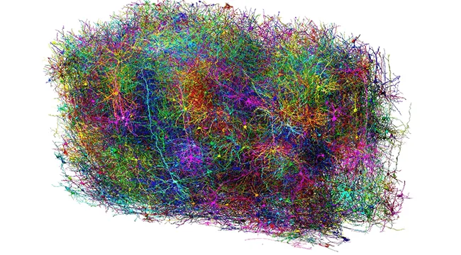
The 3D brain map includes more than 200,000 cells, 523 million synapses, and over two miles of axons, representing the most detailed wiring diagram of a piece of mammal brain ever constructed. Bock—who is associated with another project that last year unveiled the first complete map of an adult fruit fly brain—notes that the scientific advancements that led to this success bring scientists much closer to achieving the next goal: mapping an entire mouse brain.
“It’s totally doable, and I think it’s worth doing.” — Davi Bock, Ph.D.
As detailed in a collection of ten studies published Wednesday in Nature journals, the team of interdisciplinary scientists participating in the Machine Intelligence from Cortical Networks (MICrONS) project have mapped the wiring and visual functions of a piece of mouse brain roughly the size of a grain of sand. This monumental effort represents the most detailed wiring diagram of a mammal brain ever, and it holds important implications for studying brain disorders in humans. Mouse brains are similar enough to human brains that some of the things we learn from studying their neural circuitry could apply to our own. Such detailed insight into the brain’s function and structure carries important implications for understanding cognition, as well as how shifts in this wiring might be related to disorders such as Alzheimer’s, autism, Parkinson’s, and schizophrenia.