Common MRI sequences
|
|
T2 Weighted Imaging (T2) |
|
|
T1 with Fat Saturation (T1 fat-sat) |
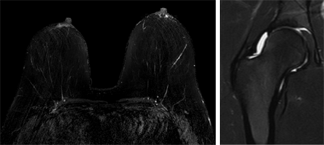
T2 with Fat Saturation (T2 fat-sat) |
|
|
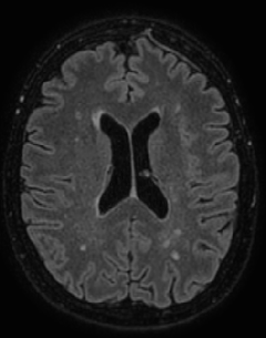
Fluid Suppression (FLAIR) |
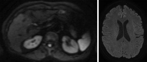
Diffusion Weighted Imaging (DWI |
|
|
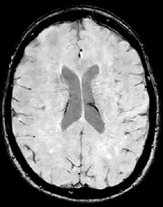
Susceptibility Weighted Imaging (SWI) |
Proton Density |
T1 with IV Gadolinium Contrast (T1-post) |
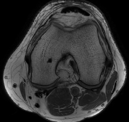
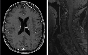
- Notice that the same tissue type (muscle, fat, abdominal organs, brain tissue, bone etc.) look different on different sequences
- Each sequence is designed to highlight different tissue types or different diseases
- There are over 100 different MRI sequences
- The simplest and most commonly used sequences are T1 and T2
Tissue representation in T1 and T2 weighted images
|
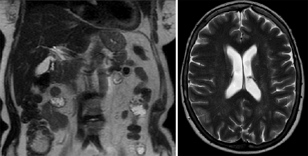
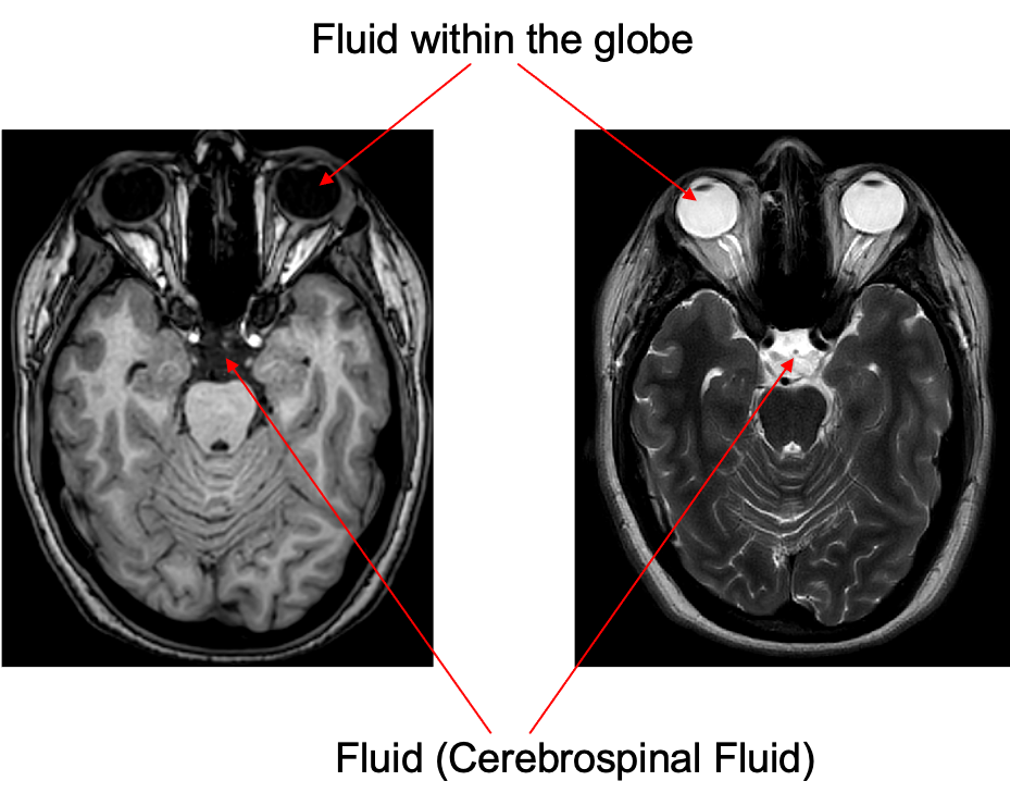
Fluid represented in a T1 and T2 weighted image |
|
|
|
|
|
T1: Dark |
T2: Bright |
|
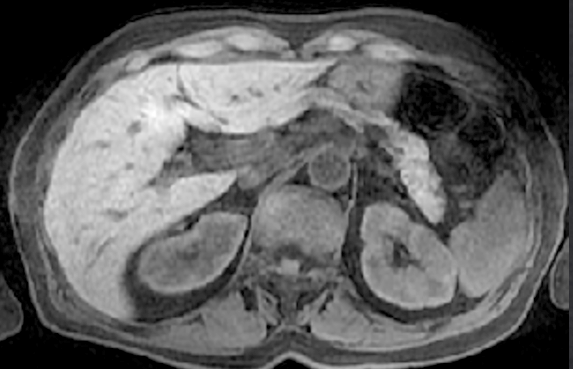
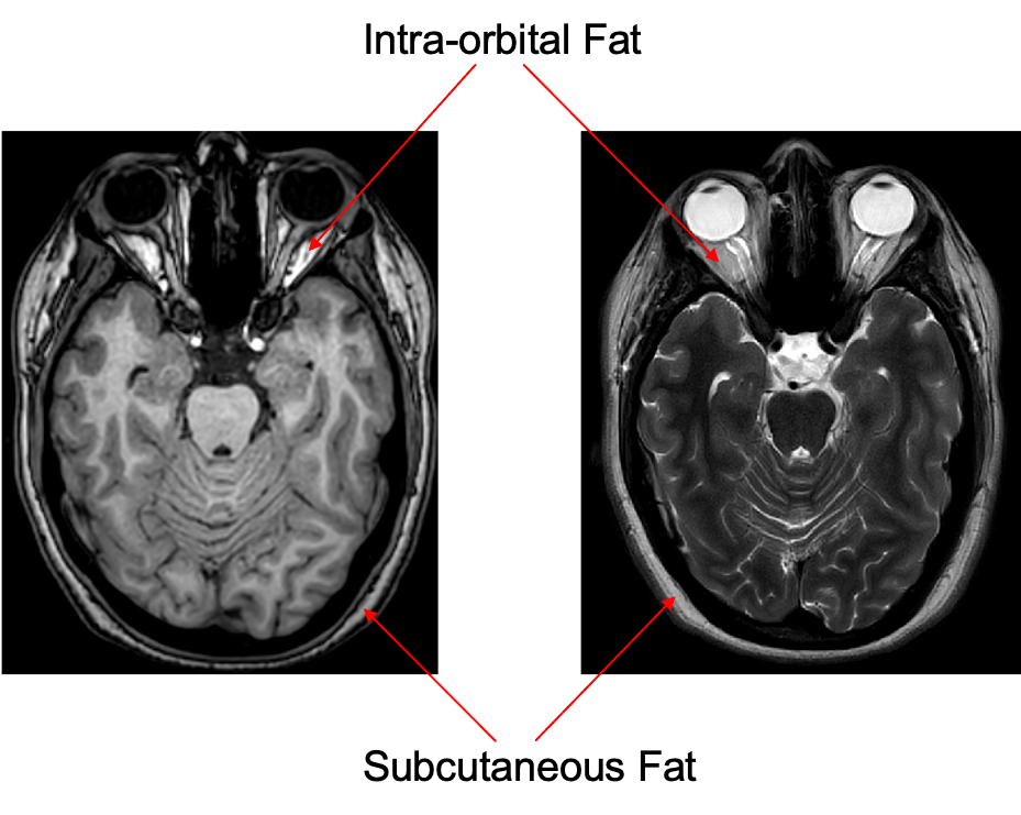
Fat represented in a T1 and T2 weighted image |
|
|
|
|
|
T1: Bright |
T2: Dark |
|
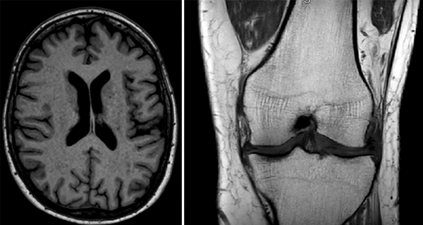
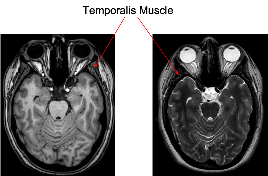
Muscle represented in a T1 and T2 weighted image. |
|
|
|
|
|
T1: Isointense |
T2: Isointens |
|
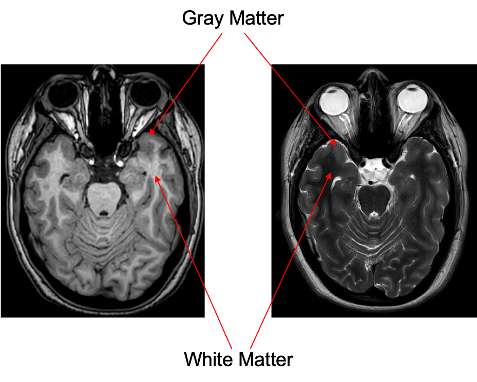
White matter represented in a T1 and T2 weighted image |
|
|
|
|
|
T1: Isointense |
T2: Isointense |
|
Signal intensity (brightness) is often described as relative signal intensity. For example:
|
|