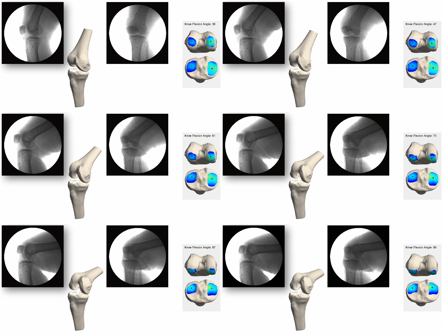
Dynamic Imaging of Joint Motion Using High-speed Dual Fluoroscopy
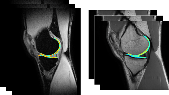
Imaging of Musculoskeletal Tissue Structure, Size and Composition
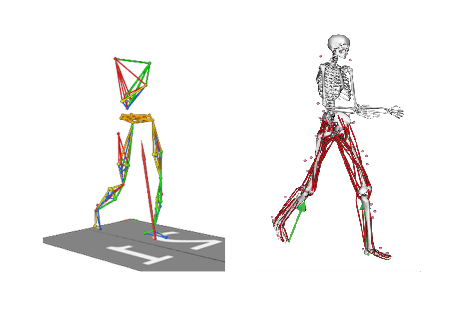
Joint Mechanics Estimation via Patient-Specific Computer Models
Improving musculoskeletal health through scientific investigations of structure and function
Dynamic Imaging of Joint Motion Using High-speed Dual Fluoroscopy
Imaging of Musculoskeletal Tissue Structure, Size and Composition
Joint Mechanics Estimation via Patient-Specific Computer Models
The overarching goal of the Musculoskeletal Imaging and Orthopaedic Biomechanics (MIOB) Laboratory is to improve human health and performance for those who suffer from musculoskeletal and orthopedic conditions.
Our approaches span experimental and computational methods. We use magnetic resonance imaging (MRI) to visualize disease and quantify anatomy, and we image bone motion directly with dynamic x-ray. Our computational studies combine patient-specific MR-based models with dynamic motion measurements to estimate internal tissue mechanics of orthopedic tissues.
Overall, the experimental and computational methods employed by the MIOB lab aim to improve health outcomes and reduce burdens imposed on patients and our healthcare system.
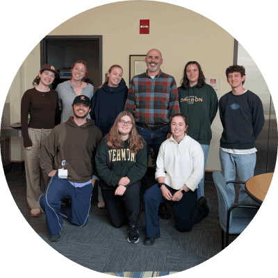
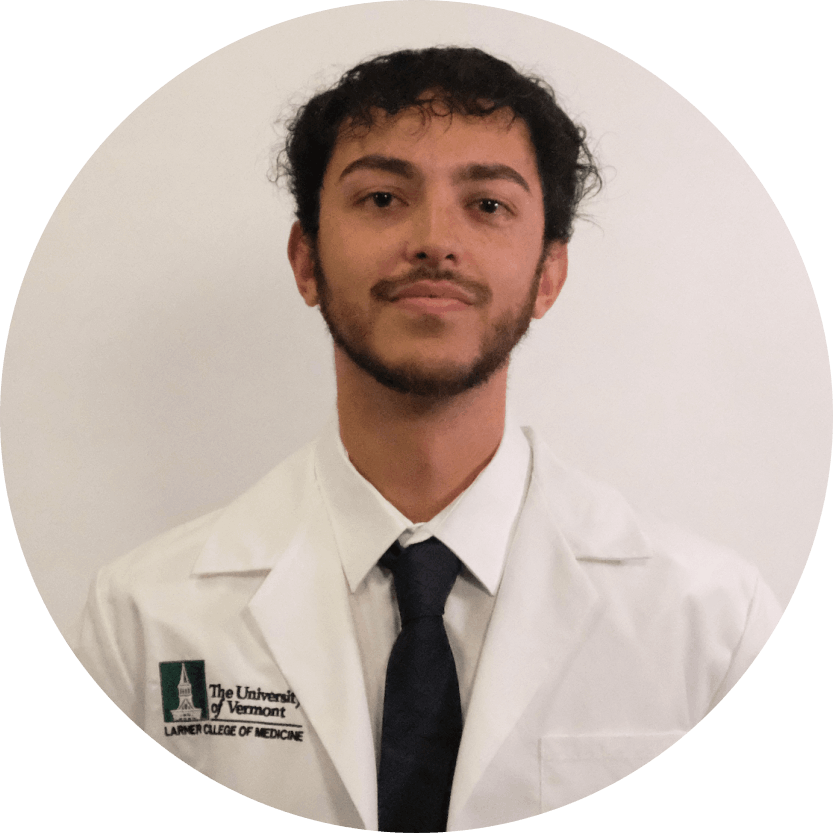
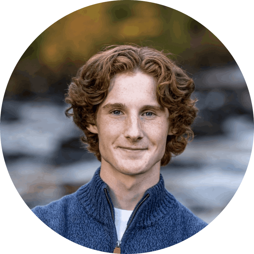
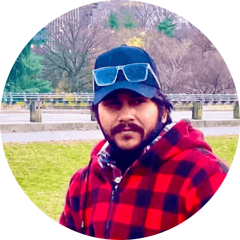
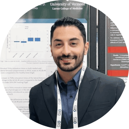
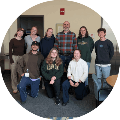 MIOB Lab
  Niccolo Fiorentino, Ph.D.
 Divya Pradip Roy, B.S.
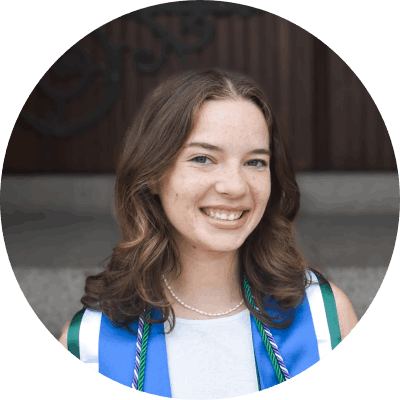 Libby Steilen, B.S.
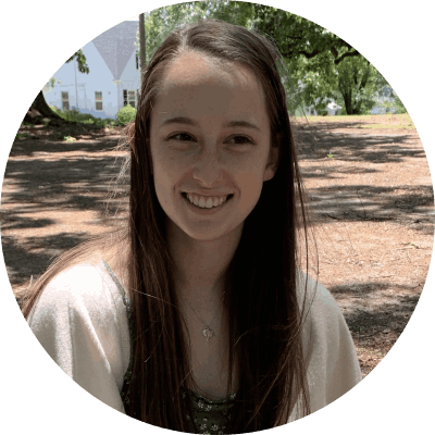 Natalie Hawley, B.S.
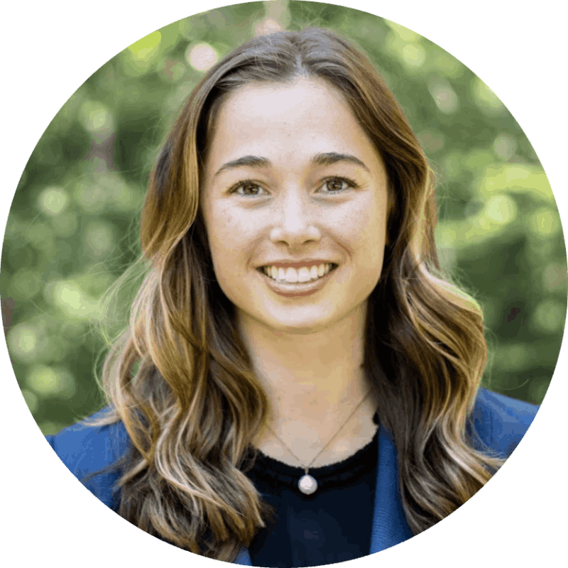 Katie Townsend, B.S.
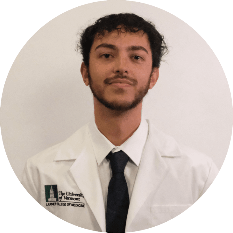 Tariq Qayum, B.S.
 Milan Shah
 Ryan Stailey
 Tasnim Sakib Apon, B.S.
 Sadegh Khodabandeloo, Ph.D., ME
 Ty Bears, B.S., BME
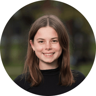 Marit Scott, M.S., BME
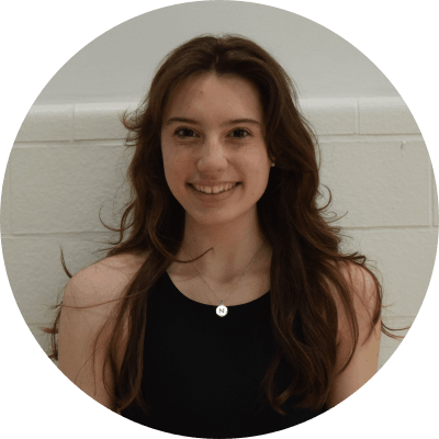 Natalie Bett, M.S.
 Julianne Boughton, M.S., BME
 Nick Taylor, B.S., BME
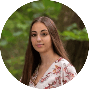 Amanda Haddad, B.S., BME
 Lincoln Lewisesquerre, B.S.
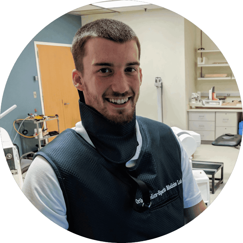 John Ramsdell, Ph.D., BME
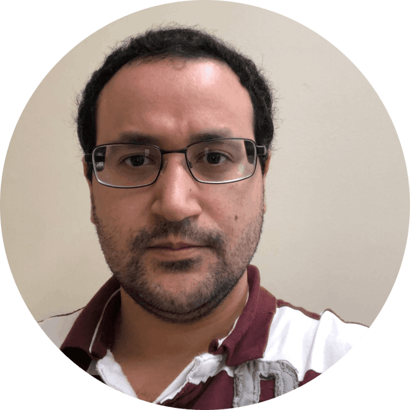 Eduardo Valdez, M.S., ME
  Haley Kuralt, M.S., BME
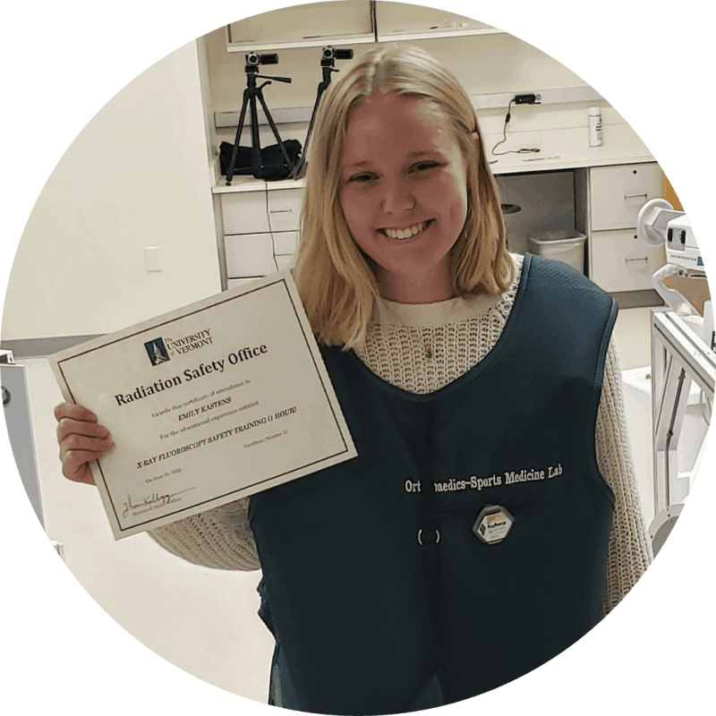 Emily Kastens, B.S., BME
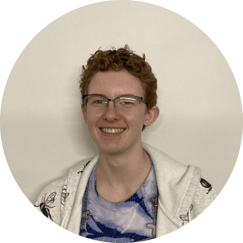 Zephyr Paxton
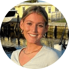 Tori Weissman, B.S., BME
  Binyameen Ghafoor, B.S., BME
  Kylie Willis, B.S., BME
  Emma Marston, B.S., BME
  James Doherty, B.S., BME
 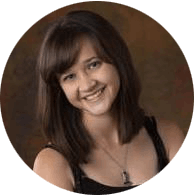 Katlyn Hall, B.S., BME
 Congratulations to Natalie H, Natalie B, and Marit on this well-deserved award. Your achievement is truly out of this world!
Sadegh graduated with his PhD in Mechanical Engineering and joined John Ramsdell in the NY Mets’ Sports Technology division. Congrats Sadegh!
Dr. Fiorentino was chosen as a 'Rising Star of Mechanical Engineering' by the American Society of Mechanical Engineering (ASME) at the 2025 International Mechanical Engineering Congress and Exposition.
Divya Pradip Roy wins UVM’s Orthopaedics Research Day Award, as voted on by researchers, staff, and clinicians at the Orthopaedics Research Day. Way to go Roy!
Dr. Fiorentino awarded the National Science Foundation's five-year Faculty Early Career Development (CAREER) grant.
Select Publications from MIOB Lab Members
Khodabandeloo S, Ramsdell JC, Beynnon BD, Krug MI, Geeslin M, Failla MJ, Choquette RH, DeSarno MJ, Fiorentino NM. Articular Cartilage Arthrokinematics and Compositional Measurements With qMRI 1 to 2 Years After ACL Reconstruction With Meniscal Surgery. Am J Sports Med. 2025 Sep;53(11):2553-2561. doi: 10.1177/03635465251360794. Epub 2025 Aug 15. PMID: 40815853
Dees A, Fiorentino NM, Krug M, DeSarno M, Vacek P, Geeslin M, Choquette R, Failla M, Zhang J, Gardner-Morse M, Beynnon B. Utilizing Quantitative MRI for Morphological Assessment of Articular Cartilage Thickness in the Tibiofemoral Joint. J Orthop Res. 2026 Feb;44(2):e70134. doi: 10.1002/jor.70134. PMID: 41606439
Scott ME, Beynnon BD, Borah AS, Ramsdell JC, Krug MI, Gardner-Morse MG, DeSarno M, Zhang J, Geeslin M, Failla MJ, Tourville TW, Fiorentino NM. Quantitative MRI-measured composition changes despite small mechanical measures in tibiofemoral cartilage of healthy adults under applied load. J Biomech. 2025 Sep;190:112864. doi.org/10.1016/j.jbiomech.2025.112864. Epub 2025 Jul 12. PMID: 40683074
Ramsdell JC, Beynnon BD, Borah AS, Gardner-Morse MG, Zhang J, Krug MI, Tourville TW, Geeslin M, Failla MJ, DeSarno M, Fiorentino NM. Tibial and femoral articular cartilage exhibit opposite outcomes for T1ρ and T2* relaxation times in response to acute compressive loading in healthy knees. J Biomech. 2024 May;169:112133. doi: 10.1016/j.jbiomech.2024.112133. Epub 2024 May 4. PMID: 38744146
Ramsdell JC, Scott ME, Beynnon BD, Fiorentino NM. Does interpolation and intra-user variability affect the accuracy of arthrokinematic measurements in the knee? A dual fluoroscopic imaging and model-based tracking study. Med Eng Phys. 2023 Apr;114:103968. doi: 10.1016/j.medengphy.2023.103968. Epub 2023 Mar 14. PMID: 37030894
Pius AK, Beynnon BD, Fiorentino N, Gardner-Morse M, Vacek PM, DeSarno M, Failla M, Slauterbeck JR, Sturnick DR, Argentieri EC, Tourville TW. Articular cartilage thickness changes differ between males and females 4 years following anterior cruciate ligament reconstruction. J Orthop Res. 2021 Jul 20. doi: 10.1002/jor.25142. Online ahead of print.
Stafford Hall
Room 411
Email: nfiorent@uvm.edu
Phone: +1 (802) 656 8082
Votey Hall
Room 231B

No Code Website Builder