1.
Images from: http://bioweb.ncsa.uiuc.edu/educwb/index2.html
2.
3.
| WHY do Sickle b globin molecules polymerize? |
In this animated
graphic, 2 molecules of sickle hemoglobin (HbS) are shown sticking
together. They stick together because the sixth
amino acid of the b-chains (blue) in HbS is
Valine (green) instead of the normal Glutamic
Acid.
| The hydrophobic
Valine side chain is exposed on the surface
of the two b-chains
of HbS and can fit into hydrophobic pockets created by the side chains
of Phenylalanine (amino
acid 85) and Leucine (amino
acid 88)
also on the
b-chain
surface.
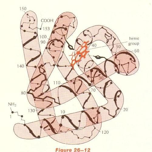
When HbS is oxygenated, the alpha helix of which valine is a part is "hinged" in position so that the valine does not reach the hydrophobic pocket. However when HbS loses its oxygen as it travels through the capillaries, the alpha helix "hinges" at a different angle, which places the 6th valine in a position where it can fit into the hydrophobic pocket. The position of amino acids 6, 85 and 88 can be seen in this drawing of a b globin chain (from Strickberger, MW. Genetics, 3rd ed., Macmillan, 1985. p 540). This does not
occur in the wild type b
globin because glutamic
acid has a very hydrophilic R group!
On the b globin graphic, find amino acids 6, 85 and 88 ! |
|
1.
Images from: http://bioweb.ncsa.uiuc.edu/educwb/index2.html
|
2. |
|
3. |
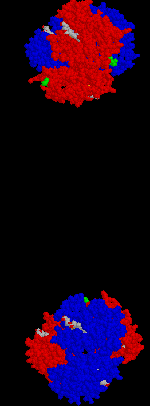
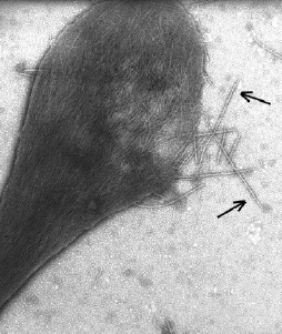
Image 1. Transmission electron micrograph (TEM) of a sickled cell showing fibers. Some of the fibers are outside of the erythrocyte. Each fiber is composed as follows:
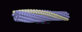
Image 2. Model of a single fiber of polymerized HbS molecules as shown in (1). The fibers in (1) are actually composed of 7 double stranded fibers twisted together - one of the double stranded fibers is shown in white.
In this image each ball represents an individual HbS tetramer. The HbS molecules are stuck together, as explained above, so that they form the double stranded fibers which then twist together to form a larger strand of 7 double strands.
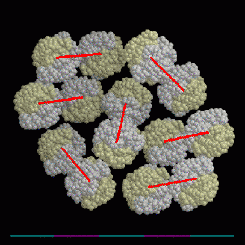
Image 3. Cross section of the large
fiber in (2). The red lines show the HbS tetramers which are paired
in each of the 7 double stranded fibers. Remember that each HbS tetramer
is composed of 2 a chains (yellow) and 2 b
chains (white). In this image the balls represent individual atoms.
| NOTE: There are many levels of structure
in the case of sickled hemoglobin
Amino Acid Sequence --> a Helix
--> a globin --> HbS tetramer
--> double stranded fiber --> 7 double stranded fibers
|
|
Primary Secondary Tertiary Quaternary 5th level 6th level Structure Structure Structure Structure |
BOTTOM LINE: A very small change in the Primary Structure of a protein may result in severe phenotypic effects! This is particularly true if the R group of the amino acid in the wild type protein is replaced by an amino acid whose R group has very different chemical properties (size, shape, solubility, charge).
WHY WOULD THIS BE?
![]()
Go to the top of the page.
| BACK to
Protein Introduction |