
The key to biochemical pathways in
living cells is catalysis of vital chemical reactions by enzymes.
Enzymes can only catalyze a chemical reaction when the shape
of its active site is complementary to the shape of its substrate.
Shown below is an example of the complementary shapes of the active
site of an enzyme (tyrosine hydroxylase) and its substrate (tyrosine):
 |
|
 |
UPPER LEFT: This is a wire frame diagram of the amino
acid tyrosine. As discussed on the previous
page, tyrosine is simply a phenylalanine
which has been hydroxylated at the para position of the phenyl ring
by phenylalanine hydroxylase. Also shown
is the amino group (the blue line represents the nitrogen atom).
On the carboxyl group, the keto oxygen is to the right, and the hydroxyl
group, with the acidic hydrogen, is to the left.
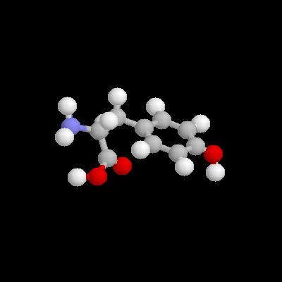
UPPER RIGHT: This is a ball and stick model of the
same tyrosine molecule, in the same orientation. The red ball at
the far right is the oxygen of the para hydroxyl group on the phenyl
ring. The amino group and the carboxyl group are readily recognizable
to the left.
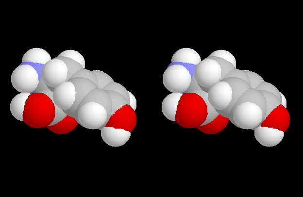
LOWER RIGHT: This is a stereo view of the same tyrosine molecule in the same orientation. To see it in 3-Dimensions, stare at it from a distance of approximately 2 feet, with your eyes unfocused. Concentrate on the middle image. When it starts to come into 3-D focus, it may help to move your head slowly in or out, or up and down slightly, or from side to side slightly. If you cannot see it in 3-D, see Dr. Barnes for a STEREOPTICON 707.
note:
tyrosine is the substrate for tyrosine
hydroxylase. The 3-Dimensional SHAPE of
this molecule gives it a complementary fit
to the active site of tyrosine hydroxylase!
LOWER LEFT: This is a stereo view of a space filling model of the active site of tyrosine hydroxylase. It is very important to see it in 3-Dimensions. Stare at it from a distance of approximately 2 feet, with your eyes unfocused. Concentrate on the middle image. When it starts to come into 3-D focus, it may help to move your head slowly in or out, or up and down slightly, or from side to side slightly. If you cannot see it in 3-D, see Dr. Barnes for a STEREOPTICON 707.
note:
the "cavity" which is formed by the active site
(the entire protein is much larger and is therefore not shown). If
you are seeing it correctly there will be a "porch roof" extending out
towards you and a "wall" on the right. The
shape of the tyrosine hydroxylase active site is complementary
to the shape of its substrate, tyrosine. When tyrosine
fits into the active site of tyrosine hydroxylase the enzyme add a second
hydroxyl group to make DOPA (dihydroxyphenylalanine).
| RETURN to "Biochemical Defect in Albinism" Page |