Details on Sickle Cell Anemia as a "molecular disease"
The human circulatory system is composed of arteries and veins.
As blood is pumped out of the heart through the aorta, there are immediately
branch arteries which carry blood to the brain, the arms and the trunk.
These arteries branch again and again and again until finally a network
of microscopic arteries is formed which contacts virtually every cell in
the body. These microscopic blood vessels are called capillary
blood vessels or simply capillaries. Their internal diameter
is just barely large enough for red blood cells to pass through single
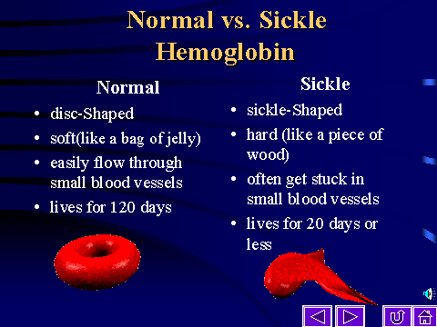
file. Red Blood Cells (erythrocytes) have a shape like a donut or a pillow.
Because they are ovoidal and flexible, they pass easily through the very
fine capillaries.
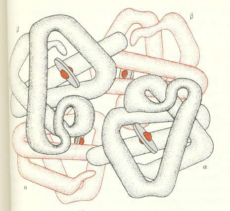
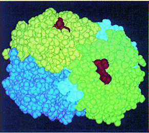
Hemoglobin is a tetrameric protein which is composed of 2 a-globin polypeptide chains and 2 b-globin polypeptide chains. This is a space-filling model of hemoglobin - with each of the 4 polypeptide chains shown in a different color. The small rust-colored ligands are the heme groups (iron atoms chelated with porphyrin rings).
As with Tay-Sachs Disease, which is just one of a larger number
of Lysosomal Storage Diseases, Sickle Cell Anemia is just one of
a large number of Hemoglobinopathies
(literally pathologies of hemoglobin).
| Recall from the discussion of the coding function of DNA that there
are a very large number of changes which can be made to a gene. For example
if only substitution mutations are considered, the gene for an average
150 amino acid protein could have 3 different alleles for each of the 450
bases - or 1,350 different alleles! Of course this also means
there there are a large number of variants for that protein!
In fact a survey of the b-globin collected from blood samples of people from cities all over the world, shows that substitutions can be found for almost every amino acid in the 146 amino acid chain:
|
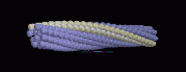
If hemoglobin contains 2 chains of sickle b-globin (HbS), the hemoglobin molecules tend to stick together because of the additional hydrophobic patches which result from the substitution of Valine for Glutamic Acid. Under anaerobic conditions, sickle cell hemoglobin - HbS - polymerizes into highly elongated cables. In the red blood cell (RBC) such polymers distort its shape and suppleness resulting in a sickle-like appearance in contrast to the normal discoid appearance of normal RBC.
The rigid sickle shape impairs the ability of the RBC to pass easily
through small capillary openings. The sickled cells become
entangled with each other and plug up the small
capillaries stopping the delivery of oxygen to the tissues in many
organs.
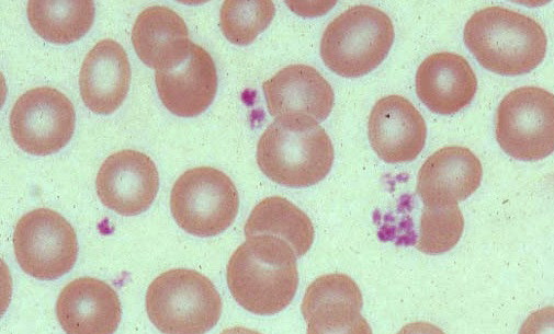
Blood smear showing normal erythrocytes. |
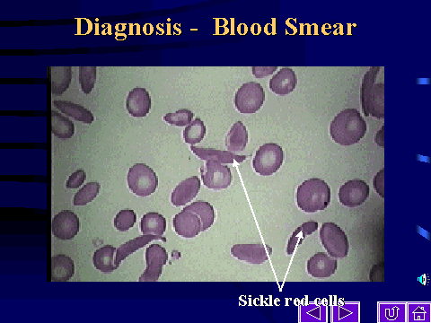
Blood smear showing sickled and normal erythrocytes. |
normal and sickled erythrocytes |
normal and sickled erythrocytes |
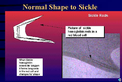
normal and sickled erythrocytes distortion of an erythrocyte by polymerizing of HbS |
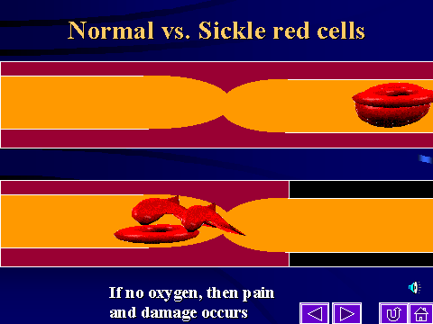
passage of normal and sickled erythrocytes throughout the capillaries. |
http://www.emory.edu/PEDS/SICKLE/prod05.htm
http://www.emory.edu/PEDS/SICKLE/tutorial/Sickle%20Cell/index.htm
Go here for more information on Sickle Cell.
PLEIOTROPIC EFFECTS
During a sickling incident many tissues and organs are damaged because their blood supply is cut off. Moreover red blood cells cannot regain their normal shape once they have become sickled. They are therefore removed from the blood supply by the spleen. This results in a severe and debilitating under supply of erythrocytes - a condition known as anemia. This in turn taxes the bone marrow which is continually stressed by an excessive demand to produce replacement red blood cells. This leads to many widespread and different types of damage, called pleiotropic effects, all of which however share a single cause - polymerization of HbS.
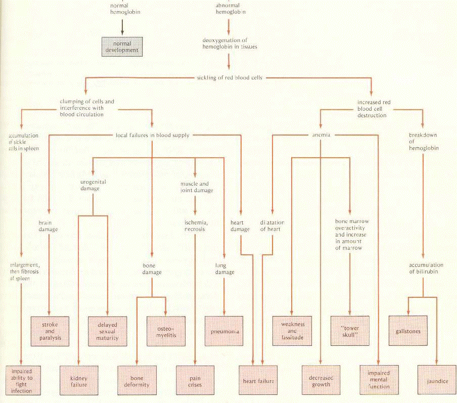 See an enlarged view of this diagram showing the pleiotropic effects.
. Strickberger, MW. Genetics, 3rd ed., Macmillan,
1985. p 543
![]()