Introduction to CHIME:
Tutorial on 3D Structure of DNA
barnes@mail.clarion.edu
Send questions or comments to Dr. Barnes!

CHIME is an application which allows you to view an organic chemical
or biochemical in 3-dimensions, from any angle. It also allows you
to display it in various ways and in different color schemes.
To see what CHIME can do, go to the CHIME
site at the University of Massachusetts.
Click on: " DNA.
An introductory level nonlinear self-paced tutorial."

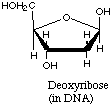
The Sugar-Phosphate
backbone
Explore the sugar-phosphate backbone by clicking on "5.
Strands and helical backbone" Now
you should see a model of the double stranded DNA molecule. Each
strand is presented in a different color. Below the model is a control
panel with "X" buttons.
-
Click on the "Y spin" button
-
Click on the "Side" button"
-
Click on "X spin"
-
Click on "Bases" and "H bonds" to erase the nitrogenous bases and hydrogen
bonds between the bases. Now we see only the sugar-phosphate backbones
of the gold and brown single strands of DNA.
-
Click on "Phosphorus". This
emphasizes how the dexoxyribose sugars are
linked together by phosphate molecules.
-
Click on "X spin".
-
Click on "Reset"
-
Click on "Bases" and "H bonds" to erase the nitrogenous bases and hydrogen
bonds between the bases.
-
Click on "Y spin". From
an end view, note how the 2 strands twist around each other to form a hollow
tube.
-
Click on "Reset"
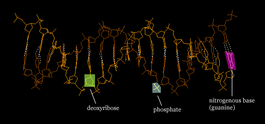
The Nitrogenous Base
Pairs
-
Click on "Sugars" to erase the deoxyribose sugars. Now you can see
the pairs of nitrogenous bases which are held together by hydrogen bonds
( white dotted lines).
-
Simultaneously move your mouse and click on the left key.
You will find that you can now manipulate the molecule in any way you choose.
Move it around so you can get a good idea of what the base pairs look like.
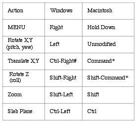
| The image can be translated
X or Y (moved left or right; up or down) with the mouse by holding
down the control key and the right button on the mouse.
The image can be yawed (rotated left and right
about the Y axis ) or pitched (rotated up or down about the X axis)
with the mouse by holding down the left button on the mouse.
The image can be rolled about the Z axis with
the mouse by holding down the shift key and the right button
of the mouse.
The image can be resized with the mouse by holding
down the shift key and the left button on the mouse. |
 |
-
Click on "Reset"
-
Click on "End"
-
Click on "Sugars" to erase the deoxyribose sugars. Now you can see
how the base pairs (held together by hydrogen bonds), form a core which
fills up the hollow center of the tube created by the sugar-phosphate backbone.
-
Click on "Reset"
A single strand of
DNA.
-
Erase the brown strand by clicking on the far left button. Now you
will see only one of the two strands of the DNA molecule.
-
Click on the buttons to erase the H bonds.
With the commands which follow, the mouse cursor
should be positioned over the black window which contains
the image. You may not get the CHIME menu otherwise.
-
Hold down the right
mouse button. On the menu which appears, go to "Select". This
brings up a sub-menu. Go to "Nucleic". This brings up a sub-menu.
Go to "DNA. We have now selected our molecule.
-
Hold down the right
mouse button. On the menu which appears, go to "Color". On
the sub-menu, go to "Monochrome". The single
strand should now appear in white.
-
Hold down the right mouse button. Choose "Select" ---> "Nucleic"
---> "Backbone"
-
Hold down the right
mouse button. On the menu which appears, go to "Color" --->
"Chain". The sugar-phosphate backbone should
now appear in blue, with the nitrogenous bases in white.
-
Click on "Phosphorus" to emphasize the links holding the deoxyribose sugars
together. Use the mouse to manipulate the model, until you have a
good idea of the way it is put together.
The Double Stranded
DNA molecule
-
Click on "Reset". Now you should see 2 strands - a blue strand and
a red strand, with the base pairs in white.
-
Click on "X spin"
-
Click on "Y spin". Now you see an end view of the molecule. Note
how the sugar-phosphate backbones form a hollow tube while the nitrogenous
base pairs fill up the core.
-
Click on "Side".
-
Use the mouse to manipulate the molecule.
-
Click on "Reset"
-
Hold down the rightmouse
button. Choose "Display" ---> "Spacefill" ---> "Van der Waals Radii".
-
Resize the molecule by holding down the shift key button. Move the mouse up or down
to zoom in or out. Reduce the molecule to the height
of your window.
-
Manipulate the molecule with your mouse to stand it up vertically.
-
Hold down the rightmouse
button. Choose "Options" ---> "Rotation".
-
Click on "X Spin".
-
Hold down the rightmouse
button. Choose "Options" ---> "Stereo Display".
| To properly see the model in stereo, put your nose
to the screen so that you can't focus on it. Slowly move back - but
do not allow your eyes to focus! If you are doing it right,
you will first see 4 spinning molecules. Concentrate on the middle
two; they will get closer together as you move farther away.
When they come together you will see the molecule in stereo! |
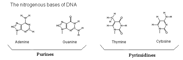
The Genetic Code
Return to the DNA Index page. This time click on the link:
"4. The Code"
This will bring up a model of double stranded DNA with the sugar-phosphate
backbones in brown, and each nucleic acid displayed in a different color.
Starting with the guanosine (green) in the lower left of the screen,
and reading toward the right ..........
-
what is the base sequence of this single strand? (Hint:
use the mouse to "roll" the molecule as you read it)
-
what is the base sequence of the complementary
single strand?
Length of Double Stranded
DNA
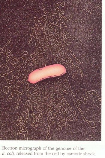
In living organisms, DNA is a very
long molecule.
-
The bacterium Escherichia coli, which
lives in our guts, has a chromosome 4,000,000 base pairs long (4 megabases).
Here is a scanning electron micrograph of a burst bacterium
with its DNA spilling out !
-
An average human chromosome has 150,000,000
base pairs (and there are 46 chromosomes in each one of our cells!).
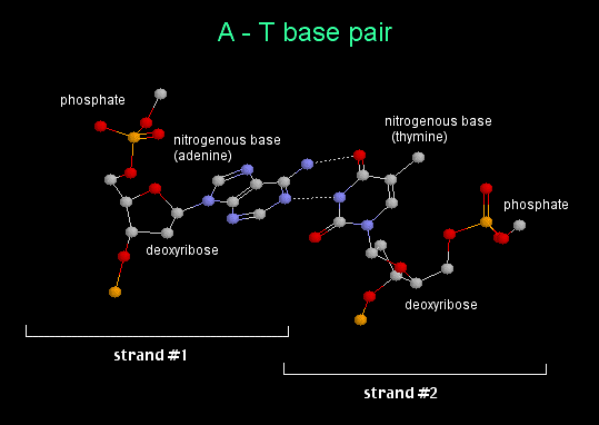
Watson and Crick
A double stranded DNA molecule is not actually shaped like a ladder as
shown in the diagram above. It is really twisted into a helix.
Imagine holding a toy ladder in your right hand. Then pointing
it away from you, twist the top with your
left hand in a
clockwise manner. This produces what is known as a
right-handed
helix.
The B-form
of DNA is a right handed helix. It is the classical structure first
described by James Watson and Francis Crick.

Go to the top
of the page.