How are Ultrasound images obtained
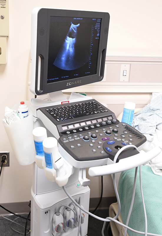
Ultrasound is so named because it uses waves above the range of human hearing (>20,000 Hz), "ultrasonic." Ultrasound uses special probes with piezoelectric quartz crystals. This means that the crystals are able to change electrical energy into mechanical energy (sound waves), send those waves into the body, and then receive waves coming back that have bounced off objects in the body, and convert them back into electrical signals which can be displayed on a screen. Ultrasound probes use the time that it takes for waves to return and the frequency of the waves to returning to create an image with different areas of light and dark.
- Transmit sound waves into the body
- Sound reflected by various tissues
- Reflected sound is captured and converted into an image
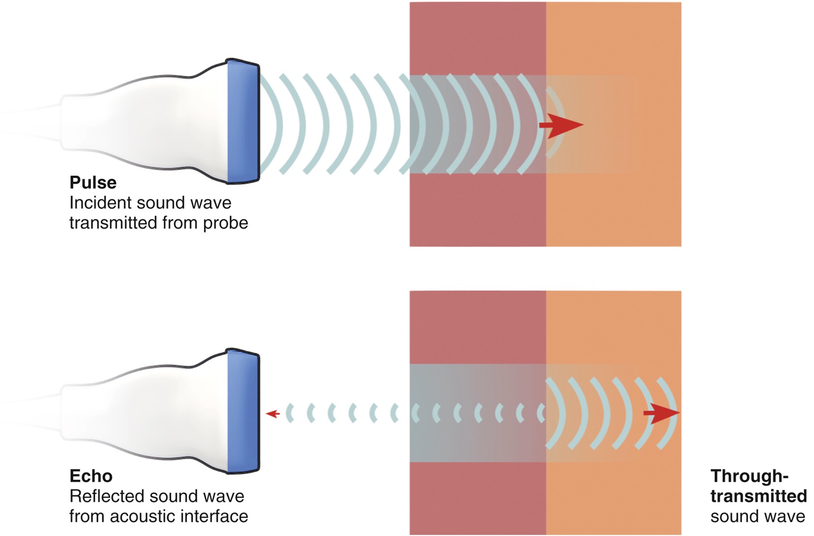
Some sound is reflected (echoed) when the ultrasound pulse encounters an interface between tissues with different values of acoustic impedence. The remaining unreflected sound energy continues on (through-transmission).
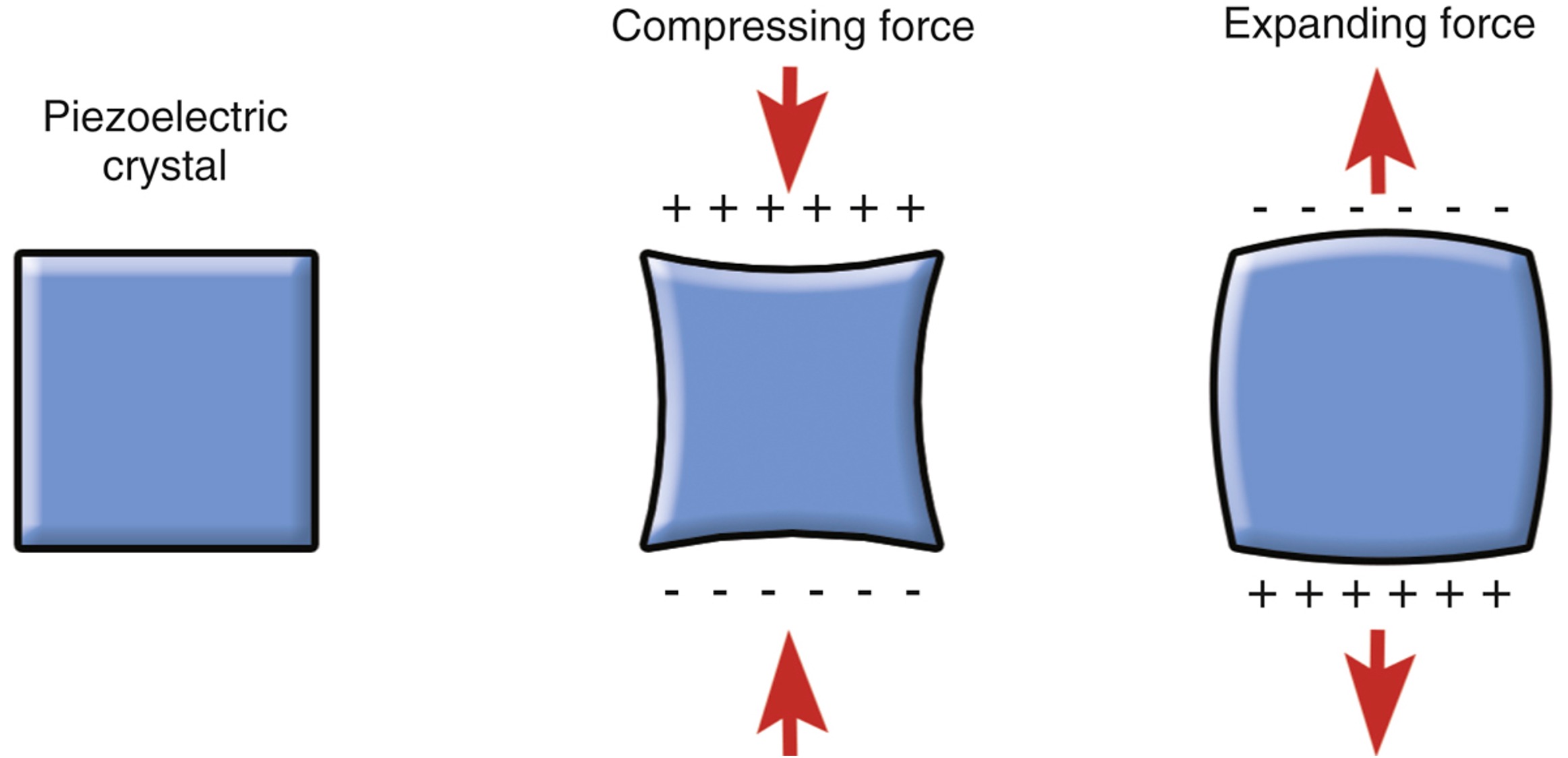
Mechanical deformation of piezoelectric crystals results in a net charge across the crystal.
How is this different from Radiographs?
- No tissue damage in the diagnostic range of frequencies
- Real-time
- Portable
- Ultrasound has no ionizing radiation that may harm the patient
The following video demonstrates the basic functionality of an ultrasound machine.
![]()