


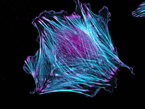

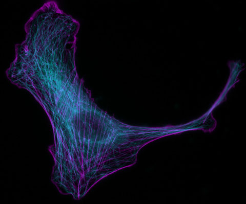


Rat embryo fibroblast stained with phalloidin (cyan) and anti-VASP antibodies (magenta)
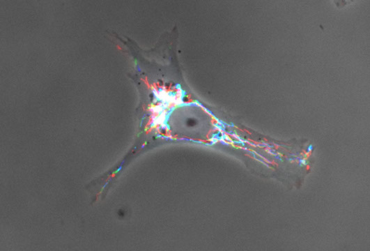
A color-coded time lapse of mitochondria infiltrating the leading edge of a migrating cell (see Cunniff & McKenzie et al, MBoC, 2016; PMID 27385336).
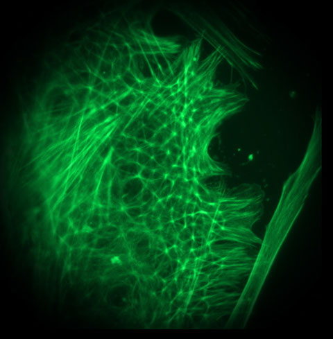
F-actin staining in a migrating rat embryo fibroblast
Mitochondrial motility in a migrating cell (see Cunniff & McKenzie et al, MBoC, 2016; PMID27385336)
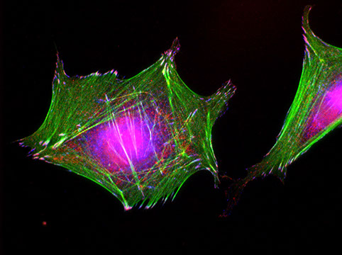
F-actin (green) and focal adhesions (red, blue) in a human dermal fibroblast
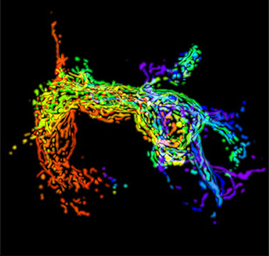
A color-coded time lapse of mitochondrial trafficking in an SKOV3ip cell invading a 3D matrix (see Cunniff & McKenzie et al, MBoC, 2016; PMID 27385336)
B16F10 mouse melanoma cell stained to visualize F-actin (cyan) & microtubules (magenta)
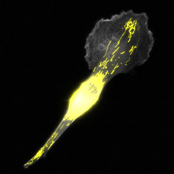
A migrating mouse melanoma cell stained to visualize the actin cytoskeleton (grey) and mitochondria (yellow)
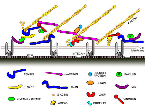
Schematic of focal adhesion proteins (modified from original image courtesy of K. Burridge, UNC-Chapel Hill)
Second-harmonic generation (SHG) imaging of the submesothelial collagen matrix of the peritoneum, a major target for ovarian cancer metastasis
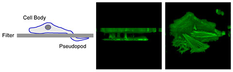
Purification of chemotactic pseudopodia - Schematic (left) and actual cell (middle & right)
Pseudopodia (cyan=F-actin; red=microtubules) protruding through two Transwell pores
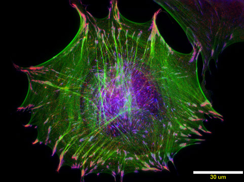
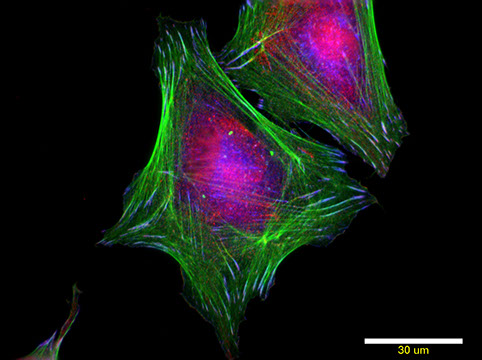
Human dermal fibroblasts (green=actin; red=VASP; blue=vinculin)
Human dermal fibroblasts treated with PDGF for 10 min (green=actin; red=VASP; blue=vinculin)
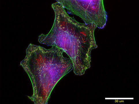
CVI-associated human dermal fibroblasts (green=actin; red=VASP; blue=vinculin)
CVI-associated human dermal fibroblasts treated with PDGF (green=actin; red=VASP; blue=vinculin)
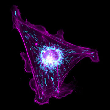
A human fibroblast stained to visualize the actin cytoskeleton (magenta) and mitochondria (cyan)
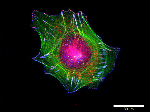
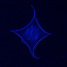
Human diploid fibroblast stained with phalloidin to visualize filamentous actin
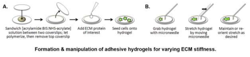
Schematic for fabrication of polyacrylamide hydrogels for investigation cellular mechanotransduction
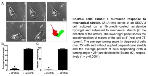
Durotaxis in human ovarian cancer cells
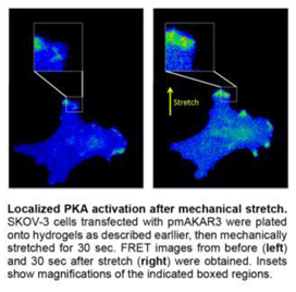
Rapid, localized activation of PKA (visualized with a FRET biosensor) in response to cellular stretch
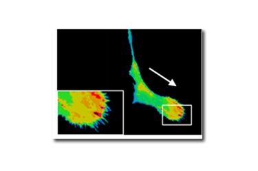
FRET image of PKA activity within the leading edge of an ovarian cancer cell invading through a 3D matrix.
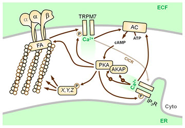
Simplified schematic for the interplay between Ca2+ and cAMP/PKA signaling
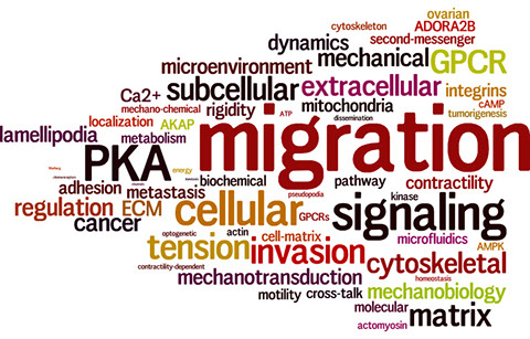
Word cloud of Howe Lab research terms
24 - 24
<
>