The Peacock Swallowtail

(Papilio Blumei)


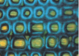
The Peacock Swallowtail is native to Indonesia and utilizes structures to produce  its colors.  The scales of this butterfly are of the type II variety.  In the previous section you compared this butterfly to the Blue Morpho and found that its color changed from green to blue when the viewing angle relative to the wings changed from straight down to almost edge on. Take another look at the specimen in the kit to verify this. The structure that produces the colors the Peacock Swallowtail presents is much different from that of the Blue Morpho, but uses some of the same physical phenomena.


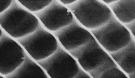
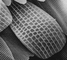
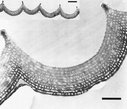
Scanning electron microscope pictures (below) of the wing reveals bowl shaped structures on the surface of the wing.  These bowls are made up of alternating layers of cuticle with pockets of air in between.  These layers are just the


right thickness to form a structure that produces  a multilayer thin film interference effect.  Recall that the tree like structures on the Blue Morpho produced this effect for the blue wavelengths of light.  But your observation of the color change with viewing angle indicates a structure that functions in a much different manner.


Take the LED flashlight  that is fitted with a polarizer and set it on top of the plastic case over part of the green stripe on one side of the butterfly. Look at this illuminated area with another piece of polaroid from as high an angle as is possible.  Rotate the polaroid until the brightness of the illuminated area is at a minimum.  What color do you see?  If the color of the wing is normally green what color is missing? 


A picture of the surface of the wing of the Peacock Swallowtail taken with an optical microscope will give you a clue as to how these structures produce color.  The blue outlines correspond to the rim
of the bowl shapes and the yellow color is from the bottom parts of the bowl.  You may want to go to the activity on polarization at this time if you are not familiar with this concept.


The polarized flashlight illuminates the bowl shaped structures with white light.  The yellow part of the light that hits the bottom of the bowls

Vukusic 2001

at little or no angle is selectively reflected back due to the spacing and index of refraction of each of the layers.  Light that hits the steeper part of the bowl is selectively reflected in the blue, because of the higher angle of the light relative to the multilayer that makes up the bowl shape.  Unlike the yellow light, the blue is not immediately reflected back out of the bowl. It is reflected to the opposite side of the bowl where it is then reflected back out.  But this extra reflection produces a change in the polarization of the light as shown in the picture to the right.
  The flat blue ribbon represents the polarized light, with the width of the ribbon representing the plane of polarization.  Notice that the reflection changes the plane of polarization.  This is why you can see the blue light but not the yellow when looking at the reflected light through a polarizer.  The butterfly wing is acting as the second polaroid in the polarization activity.  Since the yellow light is mostly reflected straight back, this bowl shape reflects mostly blue for low angles, which explains why you see mostly blue when looking at the butterfly edge on.

Vukusic 2000


Next


Vukusic 2001


Vukusic 2001


Vukusic 2001


The scale to the left is about 45 um wide. [2.4 S/D ]


Below, a closer view of the surface. The cavities are about 5 um wide. [ 22 S/D]

Below, a cross-section of the cavities showing the multilayer of about 10 layers.  The scale bar is 1um [110 S/D] and 3um   [330 S/D] (inset).