


Check out some of the lab's currently available rotation projects
Background
 Cellular interactions with the extracellular matrix (ECM) modulate nearly every major cellular behavior, including growth, division, survival and movement. A prime example of this is the concept of anchorage-dependent growth, a characteristic of most normal cells whereby division is prevented and survival is compromised in the absence of interaction with a physiologically relevant ECM. Importantly, loss of this trait is a hallmark of malignant tumor cells and correlates with their metastatic potential.
In addition to binding to & interpreting the composition of their ECM, both normal & tumor cells can sense the rigidity or pliability of the ECM. Moreover, cells dynamically react to changes in applied force or tension generated by, or in response to, ECM dynamics. Cells respond to extrinsic forces from the ECM by modifying their behavior, remodeling the ECM itself, and exerting counter-tension through actomyosin-dependent contractility.
In normal cells, this ‘mechanoreciprocity’ is in controlled equilibrium and is important for tissue homeostasis. In tumor cells, however, mechanical changes in the environment are exploited to facilitate local growth, invasion, and spread. In fact, the loss of mechanical tissue homeostasis can also often serve as a hallmark of neoplastic disease. However, the molecular mechanisms through which cells sense and respond to the mechanical nature of their ECM are not well understood. Furthermore, while the importance of microenvironmental tension for the pathogenesis of breast cancer has been elegantly established, its importance for the pathogenesis of other cancers is poorly understood.
Our Focus
 Our lab's main interest is in the mechanisms through which the extracellular micro-environment regulates cell behavior, with a specific interest in how this regulation contributes to the invasion and spread of metastatic tumors. As a postdoctoral fellow, Dr. Howe published and contributed to several articles that helped establish the field of integrin- and matrix-dependent signal transduction, in particular the regulation of MAPK activity by integrin-mediated cell adhesion. There, he also began his investigation of the role of the cAMP-dependent protein kinase (PKA) as a target for regulation by adhesion as well as a regulator of cytoskeletal dynamics and cell migration. After joining the UVM Department of Pharmacology, Dr. Howe’s lab established that, through interaction with A-kinase anchoring proteins (AKAPs), PKA is enriched within the leading edge of migrating cells and that this subcellular localization of PKA is required for efficient chemotaxis.
The laboratory’s broad efforts focus on three major questions:
- How does PKA become localized to the leading edge?
- What are the targets for PKA that are important for cell migration? and
- How does this signaling paradigm contribute to tumor cell invasion and metastasis?
To answer these questions, the laboratory combines multi-dimensional live-cell imaging, microfabrication and microfluidics, and matrix-coupled polymer hydrogels to precisely control and change the extracellular environment and to assess the molecular mechanisms used by cells to sense these changes.
Current Projects
Mechano-chemical Regulation of PKA During Cell Migration
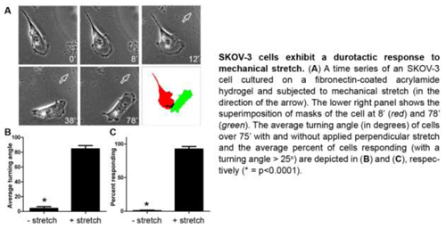
Recently, we've found that ovarian cancer (OvCA) cells exhibit durotaxis, or movement towards an increase in matrix stiffness. Moreover, when OvCA cells are mechanically stretched, PKA is rapidly and locally activated in the direction of the stretch.
 Also, PKA activation in the leading edge of migrating cells is blocked by depletion of extracellular calcium (Ca2+) and by selective inhibition of stretch-activated Ca2+ channels (SACCs). Conversely, inhibition of PKA activity or its interaction with A-kinase anchoring proteins (AKAPs) significantly reduces the frequency of SACC-mediated, tension-dependent Ca2+ transients - ‘Ca2+ flickers’ - that occur within the leading edge and are important for steering cell migration. From these observations, we hypothesize that, during cell migration, PKA activity is locally activated by intracellular tension through a mechanism that involves SACCs, and that this localized PKA activity feeds back to control Ca2+ influx. To test this hypothesis, we are currently working to determine:
- The mechanism of localized activation of PKA by mechanical stretch
- The role of stretch/tension in localized activation of PKA during cell migration
- The role of PKA in regulating Ca2+ and SACCs during cell migration
- The role of PKA, cellular tension, and SACCs in ovarian cancer metastasis in vivo
Mitochondrial Trafficking During Cell Migration and ECM Invasion
Cell migration is a complex behavior involving many energy-expensive biochemical events that iteratively alter cell shape and location. Mitochondria, the principal producers of cellular ATP, are dynamic organelles that fuse, divide, and relocate to respond to cellular metabolic demands.
 In recent work, using ovarian cancer cells as a model, we've shown that mitochondria actively infiltrate leading edge lamellipodia, thereby increasing local mitochondrial mass and relative ATP concentration and supporting a localized reversal of the Warburg shift towards aerobic glycolysis. This correlates with increased pseudopodial activity of the AMP-activated protein kinase (AMPK), a critically important cellular energy sensor and metabolic regulator. Furthermore, localized pharmacological activation of AMPK increases leading edge mitochondrial flux, ATP content, and cytoskeletal dynamics, while optogenetic inhibition of AMPK halts mitochondrial trafficking during both migration and the invasion of three-dimensional extracellular matrix. These observations indicate that AMPK couples local energy demands to subcellular targeting of mitochondria during cell migration and invasion.
Future efforts for this project include: delineation of the mechanism for AMPK-mediated mitochondrial transport; identification of the discrete cellular processes that drive metabolic flux and mitochondrial recruitment; and examination of how this energy-mediated recruitment is implemented and regulated in cells at the invasive frontier of malignant tumors.