Butternut Canker Disease
University of
Vermont Forest
Pathology
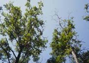
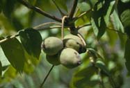 Survival of butternut (Juglans cinerea)
throughout its
range in the eastern United States and southeastern Canada is threatened by
butternut canker caused by the fungus Sirococcus
clavigignenti-juglandacearum. The severity of the disease has prompted
the United States to consider butternut a "species at risk."
Because of the rapid movement of the fungus, its extreme virulence, its lack of
genetic diveristy, and limited resistance displayed by the host, the
fungus is believed to be an exotic species of unknown origin introduced to
North America.
Survival of butternut (Juglans cinerea)
throughout its
range in the eastern United States and southeastern Canada is threatened by
butternut canker caused by the fungus Sirococcus
clavigignenti-juglandacearum. The severity of the disease has prompted
the United States to consider butternut a "species at risk."
Because of the rapid movement of the fungus, its extreme virulence, its lack of
genetic diveristy, and limited resistance displayed by the host, the
fungus is believed to be an exotic species of unknown origin introduced to
North America.
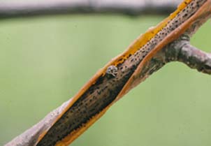
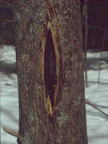
 Infection
by S. clavigignenti-juglandacearum results in
sunken, elliptical cankers on branches, stems, and buttress roots and
dark brown, elliptical stains beneath the bark. If large
cankers
(or many coalescing smaller cankers) form, branches and twigs become
girdled, resulting in dieback. The stem or buttress roots can also become
girdled, ultimately killing the tree. Trunk sprouts and epicormic shoots
usually become infected and die rapidly. Nuts have also been reported to
be infected and upon germination, seedlings may become
infected and die. Armillaria root rot is often associated with dying
trees and may hasten mortality.
Infection
by S. clavigignenti-juglandacearum results in
sunken, elliptical cankers on branches, stems, and buttress roots and
dark brown, elliptical stains beneath the bark. If large
cankers
(or many coalescing smaller cankers) form, branches and twigs become
girdled, resulting in dieback. The stem or buttress roots can also become
girdled, ultimately killing the tree. Trunk sprouts and epicormic shoots
usually become infected and die rapidly. Nuts have also been reported to
be infected and upon germination, seedlings may become
infected and die. Armillaria root rot is often associated with dying
trees and may hasten mortality.
 In Vermont, infection levels observed on 18
sites during a survey conducted from 1993 to 1995 ranged from 69 to
100%. A survey of the same trees conducted during 2001 and 2002
indicated infection rates of 71 to 96% at individual sites
and an overall infection rate of 82%. All tree locations were entered
into a database using GPS and GIS. Physical site attributes (soil type,
elevation, hydrology, aspect), which may effect disease
incidence and severity, were also obtained. To better understand the
role of each of these site factors, site attribute data will be
combined with butternut health data to determine whether there are
correlations.
In Vermont, infection levels observed on 18
sites during a survey conducted from 1993 to 1995 ranged from 69 to
100%. A survey of the same trees conducted during 2001 and 2002
indicated infection rates of 71 to 96% at individual sites
and an overall infection rate of 82%. All tree locations were entered
into a database using GPS and GIS. Physical site attributes (soil type,
elevation, hydrology, aspect), which may effect disease
incidence and severity, were also obtained. To better understand the
role of each of these site factors, site attribute data will be
combined with butternut health data to determine whether there are
correlations.
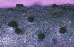
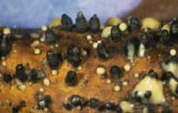 Beneath
the bark, S.
clavigignenti-juglandacearum produces thick,
black hyphal pegs or stromatal columns that cause the bark to
blister and split open. Pycnidia develop amid the hyphal pegs, and
conidiospores are released in gelatinous tendrils or creamy masses.
Spores are disseminated throughout the growing season by rain splash,
wind, and probably by insects, birds, and rodents. Fruiting
structures are more likely to develop on dead branches than on cankered
areas of the stem, and rain splash and stem runoff spread spores to lower
portions of the tree.
Beneath
the bark, S.
clavigignenti-juglandacearum produces thick,
black hyphal pegs or stromatal columns that cause the bark to
blister and split open. Pycnidia develop amid the hyphal pegs, and
conidiospores are released in gelatinous tendrils or creamy masses.
Spores are disseminated throughout the growing season by rain splash,
wind, and probably by insects, birds, and rodents. Fruiting
structures are more likely to develop on dead branches than on cankered
areas of the stem, and rain splash and stem runoff spread spores to lower
portions of the tree.
Rain splash and wind have been shown to spread conidiospores up to about
45 meters, although longer distances are possible. Since its discovery
in 1967, the fungus has spread rapidly and efficiently throughout the
range of butternut, raising questions about the mode of
dissemination. Vectors specifically targeting butternut trees may be
involved. Sticky conidiospores could adhere easily to the
exoskeleton of winged insects and be transported long distances.
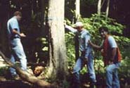
And so began our search for potential insect
vectors of the butternut canker fungus in Vermont.
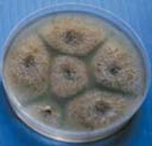 Sirococcus clavigignenti-juglandacearum is easily isolated from the
margins of cankers onto malt extract agar. At 20 degrees Celsius in the
dark,
the fungus produces brown to black pycnidia in about 14 days.
Sirococcus clavigignenti-juglandacearum is easily isolated from the
margins of cankers onto malt extract agar. At 20 degrees Celsius in the
dark,
the fungus produces brown to black pycnidia in about 14 days.
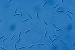
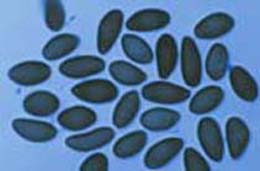
Conidiospores are
hyaline (clear),
fusiform (spindle-shaped), 2-celled, and measure 9-17 micrometers x 1-1.5
micrometers. A perfect stage of the
fungus remains unknown.
 A
secondary fungus, Melanconium oblongum (perfect
stage: Melanconis juglandis), is often found fruiting on dead
butternut branches and is often confused with S.
clavigignenti-juglandacearum. Melanconium oblongum produces
black
acervuli and ovoid to ellipsoid, 1-celled, dark conidiospores that average
19 micrometers x 9 micrometers.
A
secondary fungus, Melanconium oblongum (perfect
stage: Melanconis juglandis), is often found fruiting on dead
butternut branches and is often confused with S.
clavigignenti-juglandacearum. Melanconium oblongum produces
black
acervuli and ovoid to ellipsoid, 1-celled, dark conidiospores that average
19 micrometers x 9 micrometers.
Considered a weak parasite, this fungus invades weakened
or dead tissue and causes what is referred to as Melanconis dieback.
Both S. clavigignenti-juglandacearum and M. oblongum can be
found fruiting on the same branch.
Search for
Potential Insect
Vectors of
the Butternut Canker Fungus in Vermont
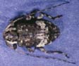
 |
Acoptus suturalis |
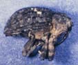
 |
Astylopsis macula |
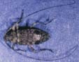
 |
Eubulus parochus |
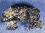
 |
Hyperplatys maculata |
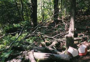
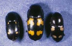
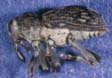
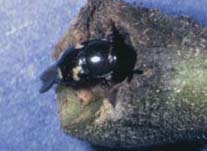
From 1997 to 1999, we found at least 17 species of
beetles (Coleoptera), representing 8
families, carrying conidiospores of Sirococcus
clavigignenti-juglandacearum at two sites in northern Vermont. Most
species belonged to the families Cerambycidae (longhorned beetles) and
Curculionidae (weevils).
Total numbers of conidiospores estimated per
beetle ranged
from 200 to 1.6 million. Beetles found in greatest abundance from freshly
cut logs and branches of butternut upon which the fungus was fruiting
included: Acoptus suturalis (Curculionidae), Astylopsis
macula (Cerambycidae), Eubulus parochus (Curculionidae), and
Hyperplatys maculata (Cerambycidae).
In 1999, 37 to 74% of each of
these four species was carrying conidiospores of the butternut canker
fungus. We observed these beetles feeding on hypal pegs
and pycnidia of the fungus.
They were also found in crowns of living trees where they
are probably attracted to dead tissue. They may be carrying spores of
S. clavigignenti-juglandacearum to both
living and recently dead branches and increasing infections in the
crowns.
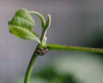
Beetles
most commonly collected from crowns of live butternut trees
included: the butternut curculio, Conotrachelus juglandis
(Curculionidae) and a leaf beetle, Paria sp. (Chrysomelidae). The
butternut curculio creates feeding and egg-laying wounds on living shoots.
From 1997 to 1999, 6-11% of curculios carried conidiospores. Although the
numbers they carried were relatively small, it is possible these spores
would be sufficient to infect healthy or freshly wounded tissue of
butternut. Curculio wounds may make suitable infection courts for
conidiospores transported by rain splash or insects.
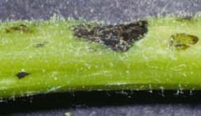
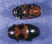
 We collected several
species of sap-feeding beetles (Nitidulidae), a small
percentage of which carried conidiospores of S.
clavigignenti-juglandacearum. We observed nitidulids crawling in
oozing butternut cankers and burrowing into curculio wounds on living
shoots. Nitidulids could be important vectors of the fungus if they move
from sticky, sporulating structures to wounds made by the curculio or
other agents on live shoots and branches of butternut.
We collected several
species of sap-feeding beetles (Nitidulidae), a small
percentage of which carried conidiospores of S.
clavigignenti-juglandacearum. We observed nitidulids crawling in
oozing butternut cankers and burrowing into curculio wounds on living
shoots. Nitidulids could be important vectors of the fungus if they move
from sticky, sporulating structures to wounds made by the curculio or
other agents on live shoots and branches of butternut.
If you would like to find out more, see our publication:
Halik, S. and Bergdahl, D.R. 2002. Potential beetle vectors of
Sirococcus clavigignenti-juglandacearum on butternut. Plant
Disease 86:521-527.